What Are The 2 Types Of Microscopes?
The Two Major Types of Microscopes: A Detailed Overview
Microscopy is an essential tool in the world of science, helping researchers explore the microscopic world and understand the fine details of various biological and material structures. There are numerous types of microscopes, but broadly speaking, they can be classified into two major categories: optical microscopes and electron microscopes. Each type operates on distinct principles and is suited for different applications, but both play a crucial role in advancing scientific knowledge.
1. Optical Microscopes: The Classic Workhorse
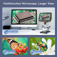
Optical microscopes, also known as light microscopes, are the most common and widely used type. They use visible light to illuminate specimens and a system of lenses to magnify the image. They have been the cornerstone of biology and medicine for centuries, and even with advances in technology, they remain indispensable in laboratories around the world.
Basic Principle

Optical microscopes work by passing visible light through a specimen. The light is then focused by lenses, typically made of glass or sometimes plastic, to form an image of the object being viewed. The lenses bend or "refract" light to magnify the object, and the image is projected onto the viewer's eyes or captured by a camera.
Types of Optical Microscopes

1. Compound Microscopes:
Compound microscopes are the most common type of optical microscope. They have multiple lenses to provide high magnification and are generally used to observe thin slices of specimens, such as cells, tissues, or microorganisms.
- Structure: A compound microscope typically has an objective lens (which is closest to the specimen) and an eyepiece lens (the lens through which the user views the image). The objective lenses often come in several magnification powers, such as 4x, 10x, 40x, and 100x.
- Magnification: Depending on the combination of objective lenses and eyepiece, magnification typically ranges from 40x to 1,000x.
- Applications: These microscopes are used extensively in biology, medical diagnostics, and material science to observe cells, bacteria, tissues, and other small structures.
2. Stereo (Dissecting) Microscopes:
Unlike compound microscopes, stereo microscopes provide a three-dimensional view of a specimen. They are often used for larger objects and provide a lower magnification.
- Structure: These microscopes typically have two eyepieces and two separate light paths. The light is reflected off the specimen from above, allowing the user to see the surface features of larger specimens in three dimensions.
- Magnification: Stereo microscopes usually offer magnification ranges from 10x to 100x, which is much lower than that of compound microscopes but ideal for examining larger structures like insects, circuit boards, or geological samples.
- Applications: They're commonly used in fields like electronics, material science, and entomology for inspecting larger specimens in detail.
Advantages of Optical Microscopes

- Simplicity: They are relatively simple to use, which makes them accessible to both professionals and beginners.
- Cost-Effective: Optical microscopes are far more affordable than electron microscopes, making them ideal for most educational and clinical settings.
- Real-time Viewing: Since they use visible light, optical microscopes allow users to observe specimens in real-time, which is critical for certain live-cell studies or dynamic processes.
Limitations of Optical Microscopes

- Resolution: Optical microscopes are limited by the wavelength of light, which is typically around 400 to 700 nanometers. This means they cannot resolve objects smaller than roughly 200 nanometers, limiting their ability to observe subcellular structures or smaller components.
- Depth of Field: Although optical microscopes are great for viewing thin sections of a specimen, they generally lack the depth of field needed to observe complex, three-dimensional structures in full.
2. Electron Microscopes: Unlocking the Nano-World
Electron microscopes (EM) represent a giant leap forward in microscopy, offering unparalleled resolution. Unlike optical microscopes, which use visible light, electron microscopes use a beam of electrons to illuminate specimens. Electrons have much shorter wavelengths than visible light, allowing electron microscopes to achieve much higher magnifications and resolutions.
Basic Principle
Electron microscopes work by firing a beam of electrons at a specimen. The electrons interact with the atoms in the specimen, and this interaction produces signals that can be used to generate an image. These microscopes rely on electromagnetic lenses to focus the electron beam instead of optical lenses, which enables them to resolve much smaller structures.
Types of Electron Microscopes
1. Transmission Electron Microscopes (TEM):
TEMs are capable of achieving the highest resolution among all types of microscopes, making them invaluable for studying the internal structure of cells, viruses, and other extremely small specimens.
- Structure: A TEM uses a very thin section of the specimen, often just one or two molecules thick, which is bombarded with electrons. The electrons pass through the specimen, and the resulting image is captured on a phosphorescent screen or digital detector.
- Magnification: TEMs can provide magnifications up to 10 million times and resolutions down to the atomic level, far surpassing the limits of optical microscopes.
- Applications: TEMs are primarily used in materials science, nanotechnology, and cellular biology. They are essential tools for observing viruses, bacteria, and even the internal structures of organelles within cells.
2. Scanning Electron Microscopes (SEM):
SEMs differ from TEMs in that they scan the surface of a specimen with a focused electron beam. Rather than passing electrons through a sample, SEMs bounce the electrons off the surface and detect the reflected signals to generate an image.
- Structure: SEMs have a specimen that is usually coated with a thin layer of conductive material, such as gold or platinum, to prevent charging effects from the electron beam.
- Magnification: SEMs typically offer magnifications from 10x to 500,000x, with resolution on the order of 1 nanometer.
- Applications: SEMs are commonly used in industrial applications, material sciences, and for detailed surface analysis of objects like metals, composites, and biological tissues.
Advantages of Electron Microscopes
- Extreme Resolution: The biggest advantage of electron microscopes is their ability to observe specimens at a much higher resolution than optical microscopes, revealing fine details like molecular structures, viruses, and nanoparticles.
- 3D Imaging (SEM): While TEM offers detailed internal views, SEM provides detailed three-dimensional surface images, making it ideal for studying surface topography and morphology.
Limitations of Electron Microscopes
- Cost and Size: Electron microscopes are significantly more expensive and larger than optical microscopes, often requiring specialized rooms and maintenance.
- Sample Preparation: Electron microscopy requires specimens to be prepared in very specific ways, including dehydration, thin sectioning (for TEM), and coating with conductive materials (for SEM). This can introduce artifacts or distortions in the image.
- No Live Samples: Electron microscopes generally cannot observe live specimens because the sample preparation process and the vacuum environment are incompatible with living organisms.
Comparing Optical and Electron Microscopes
| Feature | Optical Microscopes | Electron Microscopes |
|-------------------------------|-----------------------------------|------------------------------------|
| Principle | Uses light to illuminate samples | Uses electron beams to illuminate samples |
| Resolution | ~200 nm (limited by light wavelength) | Sub-nanometer resolution (down to atomic level) |
| Magnification | 40x to 1,000x | Up to 10 million times (TEM), 500,000x (SEM) |
| Live Sample Observation | Yes | No |
| Cost | Relatively affordable | Expensive, often requiring specialized facilities |
| Sample Preparation | Simple | Complex, often requires coating, dehydration, etc. |
In conclusion, both optical microscopes and electron microscopes are indispensable tools in modern science, each serving different purposes and offering unique advantages. Optical microscopes are accessible, easy to use, and ideal for everyday tasks like observing living cells or tissue samples. On the other hand, electron microscopes provide an incredible level of detail, revealing the tiniest structures in the world, but at a much higher cost and complexity. By understanding the strengths and limitations of each type of microscope, researchers can choose the right tool for their specific needs, ensuring more accurate and informative observations.


There are no comments for this blog.