What Is The Field Of View Microscope?
Microscopes are indispensable tools in scientific research, clinical laboratories, and educational settings. Among the various technical terms and parameters that define a microscope's capabilities, the "field of view" (FOV) is one of the most critical. This article delves into the concept of the field of view in microscopy, its significance, factors affecting it, and provides guidance on optimizing it for various applications.
Understanding the Field of View
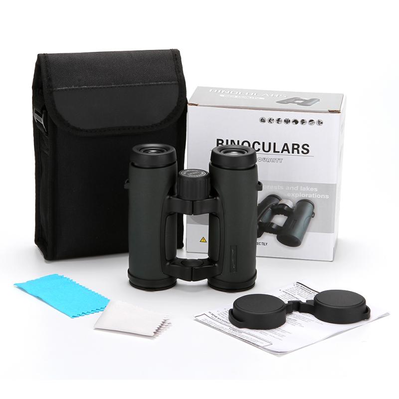
The field of view of a microscope refers to the extent of the observable world that can be seen at any given moment through the microscope's eyepiece or camera. Essentially, it is the circle of light visible when one looks through the microscope. The dimension of this circle is usually measured in micrometers (µm) or millimeters (mm) and varies depending on the magnification settings and the specific microscope design.
Importance of the Field of View
1. Image Clarity and Context:
- A larger field of view allows the observer to see more of the specimen within a single glance, which can provide greater context and make it easier to locate specific structures or features.
2. Efficiency in Observations:
- When scanning large specimens, a larger field of view reduces the need to move the slide as frequently, thus speeding up the observation process.
3. Quantitative Analysis:
- In research involving cell counting, spatial distribution, or tissue mapping, an appropriate field of view is crucial for accurate quantitative analysis.
Factors Affecting the Field of View
1. Objective Lens Magnification:
- Higher magnifications result in a smaller field of view. For instance, a 10x objective lens will have a larger field of view compared to a 40x lens.
2. Eyepiece Lens:
- The design and specification of the eyepiece can influence the field of view. Wide-field eyepieces are designed to offer a larger field of view for the same magnification compared to standard eyepieces.
3. Tube Length and Design:
- The physical dimensions and the internal design of the microscope's optical tube can also impact the overall field of view.
4. Digital Camera Sensor Size (in digital microscopy):
- When using digital cameras, the sensor size of the camera influences the field of view. Larger sensors typically provide a broader field of view.
How to Measure the Field of View
The process involves using a stage micrometer, which is a special slide marked with an accurate scale in micrometers. Place the micrometer slide on the microscope stage, focus, and count the divisions visible within the field of view.
1. Select the objective lens to be used.
2. Place the stage micrometer on the microscope stage and bring it into focus.
3. Count the number of divisions visible across the diameter of the circle of light.
4. Multiply the number of divisions by the micrometer scale (usually given on the micrometer slide).
Calculating Field of View
The field of view can be approximately calculated using a formula:
\[ FOV_{\text{new}} = \frac{FOV_{\text{old}} \times \text{Total Magnification}_{\text{old}}}{\text{Total Magnification}_{\text{new}}} \]
Where:
- \( FOV_{\text{new}} \) is the field of view with the new magnification.
- \( FOV_{\text{old}} \) is the known field of view with the old magnification.
- \( \text{Total Magnification}_{\text{old}} \) is the total magnification using the old setup.
- \( \text{Total Magnification}_{\text{new}} \) is the total magnification using the new setup.
Optimizing Field of View for Specific Applications
1. Clinical Diagnostics:
- Pathologists often require a balance between high magnification and sufficient field of view to observe tissue samples effectively.
2. Educational Purposes:
- Wide-field eyepieces are typically advantageous in educational settings to allow students to easily locate and study specimens.
3. Research Applications:
- Research applications may demand either a broader field of view or higher magnification with a narrower field of view depending on the specifics of the study. Researchers should select objectives and eyepieces that optimize their workflow.
4. Photography and Digital Documentation:
- For microscopic imaging, the camera's sensor size should be balanced with the microscope's optics to capture the maximum area without compromising image resolution.
Common Challenges and Solutions
1. Narrow Field of View at High Magnifications:
- This is a common challenge which can be mitigated by using wide-field eyepieces or by stitching multiple images together in software to create a composite view.
2. Edge Distortion:
- Some microscopes might show distortion at the edges of the field of view. This can often be minimized by using flat-field objectives and ensuring proper alignment of optical components.
Understanding and optimizing the field of view in microscopy is essential for effective observation, accurate data collection, and efficient workflow. Whether you are a researcher, clinician, or educator, grasping the intricacies of the field of view can greatly enhance the quality and accuracy of your microscopic examinations. By considering factors such as objective lens magnification, eyepiece design, and additional digital equipment, you can tailor your microscope setup to meet specific needs and achieve the best possible visual results.




















There are no comments for this blog.